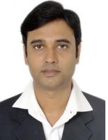
Dr. Vishal Srivastava
Professor
Specialization
Deep Learning for Healthcare Devices
vsrivastava@thapar.edu
Specialization
Deep Learning for Healthcare Devices
Biography
Dr. Vishal Srivastava received his Ph.D. and Master's degree in Instrumentation from Indian Institute of Technology Delhi. He was a post-doctoral fellow in the Biomedical Engineering Department at the University of Huston, USA and in the Electrical and Computer Engineering Department at University of California Los Angles, USA. He is an Associate Editor of IEEE Access and various other journals. He has more than 30 publications in the SCI journals. He has supervised two PhD students and 3 are undergoing.
| Phone/Mobile Number | 9990435475 |
| Email ID | vsrivastava@thapar.edu |
Honors and awards
- Post-Doctoral fellow in the Electrical and Computer Engineering Department, University of California Los Angeles, USA.
- Post-Doctoral fellow at Department of Biomedical Engineering, University of Houston, USA.
- Associate Editor of IEEE Access Journal.
Service activities (within and outside of the institution)
- Time Table in charge, UG Coordinator of Biomedical Engineering
Publications:
- Sharma et al., "Machine learning for forecasting the biomechanical behavior of orthopedic bone plates fabricated by fused deposition modelling," Rapid Prototyping J., 2024.
- Dhiman, S. Kamboj, and V. Srivastava, "Explainable AI based efficient ensemble model for breast cancer classification using optical coherence tomography," Biomed. Signal Process. Control, vol. 91, p. 106007, 2024.
- Sharma et al., "Optimization of polydopamine coating process for poly lactic acid-based 3D printed bone plates using machine learning approaches," Polym. Eng. Sci., vol. 64, no. 1, pp. 279-295, 2024.
- Sharma et al., "Predicting biomechanical properties of additively manufactured polydopamine coated poly lactic acid bone plates using deep learning," Eng. Appl. Artif. Intell., vol. 124, p. 106587, 2023.
- Basu, R. Agarwal, and V. Srivastava, "Development of an Intelligent Full‐Field Polarization Sensitive Optical Coherence Tomography for Breast Cancer Classification," J. Biophotonics, p. e202200385, 2023.
- Basu, R. Agarwal, and V. Srivastava, "Deep discriminative learning model with calibrated attention map for the automated diagnosis of diffuse large B-cell lymphoma," Biomed. Signal Process. Control, vol. 76, 2022.
- Kansal, J. Bhattacharya, and V. Srivastava, "Automated full-field polarization sensitive optical coherence tomography diagnostic systems for breast cancer," Appl. Opt., vol. 59, pp. 7688-7693, 2020.
- Singla and V. Srivastava, "Deep learning enabled multi-wavelength spatial coherence microscope for the classification of malaria-infected stages with limited labelled data size," Opt. Laser Technol., vol. 130, p. 106335, 2020.
- Singh, V. Srivastava, and D. S. Mehta, "Machine learning-based screening of red blood cells using quantitative phase imaging with micro-spectrocolorimeter," Opt. Laser Technol., vol. 124, p. 105980, 2020.
- Dubey et al., "Self-attention based BiLSTM-CNN classifier for the prediction of ischemic and non-ischemic cardiomyopathy," Laser Phys. Lett., vol. 17, 2020.
- Kansal, S. Goel, J. Bhattacharya, and V. Srivastava, "Generative adversarial network–convolution neural network based breast cancer classification using optical coherence tomographic images," Laser Phys., vol. 30, p. 115601, 2020.
- Singla, V. Srivastava, and D. S. Mehta, "Development of full-field optical spatial coherence tomography system for automated identification of malaria using the multilevel ensemble classifier," J. Biophotonics, vol. 11, no. 5, 2018.
- Dubey, V. Srivastava, and K. Dalal, "In Vivo Automated Quantification of Thermally Damaged Human Tissue Using Polarization Sensitive Optical Coherence Tomography," Comput. Med. Imaging Graph., vol. 64, 2018.
- Gaur et al., "Fire Sensing Technologies: A Review," IEEE Sensors, 2019.
- Verma et al., "Sensing, Controlling, and IoT Infrastructure in Smart Building: A Review," IEEE Sensors, vol. 19, no. 20, 2020.
- Singla and V. Srivastava, "Deep learning enabled multi-wavelength spatial coherence microscope for the classification of malaria-infected stages with limited labelled data size," Opt. Laser Technol., vol. 130, p. 106335, 2020.
- Tulsi Anna, Sandeep Chakraborty, Chia-Yi Cheng, Vishal Srivastava, Arthur Chiou, Wen-Chuan Kuo, “Elucidation of microstructural changes in leaves during senescence using spectral domain optical coherence tomography” , Scientific reports, (Nature Group), 9, (2019).
- Neeru Singla, Kavita Dubey, Vishal Srivastava, “Automated assessment of breast cancer margin in optical coherence tomography images via pre-trained convolutional neural network", Journal of Biophotonics, Vol. 12, 3, (2019).
- Anshul Gaur, Abhishek Singh, Ashok Kumar, Kishor S Kulkarni, Sayantani Lala, Kamal Kapoor, Vishal Srivastava, Anuj Kumar, SC Mukhopadhyay, “Fire Sensing Technologies: A Review”, IEEE Sensors (2019).
- Anurag Verma, Surya Prakash, Vishal Srivastava, Anuj Kumar, and S. C. Mukhopadhyay, “Sensing, Controlling, and IoT Infrastructure in Smart Building: A Review”, Vol. 19, Issue 20, IEEE Sensors (2020).
- Neeru Singla, and Vishal Srivastava, “Deep learning enabled multi-wavelength spatial coherence microscope for the classification of malaria-infected stages with limited labelled data size” Vol 130, Page no 106335, Optics & Laser Technology (2020).
- Neeru Singla, Vishal Srivastava, Dalip Singh Mehta, “Development of full-field optical spatial coherence tomography system for automated identification of malaria using the multilevel ensemble classifier", Journal of Biophotonics, Vol. 11, 5, (2018).
- Kavita Dubey, Vishal Srivastava, and Krishna Dalal, "In Vivo Automated Quantification of Thermally Damaged Human Tissue Using Polarization Sensitive Optical Coherence Tomography ", Computerized Medical Imaging and Graphics, Vol. 64, (2018).
- Vishal Srivastava, Sreyankar Nandy and Dalip Singh Mehta, “High-resolution full-field optical coherence tomography using a spatially incoherent monochromatic light source”, Applied Physics letter, Vol. 103, 103702 (2013).
- Vishal Srivastava, Sreyankar Nandy and Dalip Singh Mehta, “High-resolution corneal topography and tomography of fish eye using wide field white light interferenc microscopy”, Applied Physics letter, Vol. 102, 153701 (2013).
- Dalip Singh Mehta and Vishal Srivastava, “Quantitative phase imaging of human red blood cells using phase-shifting white light interference microscopy with colour fringe analysis”, Applied Physics letter, Vol.101, 203701 (2012).
Sponsored Project
- “Intelligent 3D microscope for biological cells classification,” Supervised by (PI/CoPI) received Grant amount of Rs 45.73 lacs by SERB under CRG scheme for the duration of March 2022 to March 2025.
- “Health Care System: Early Detection of Bone Cancer using Microwave Imaging and Treatment by Hyperthermia Lens Applicator,” Supervised by (PI/CoPI) received Grant amount of Rs 52.70 lacs by SERB under CRG scheme for the duration of Sept. 2023 to Sept. 2026.
- “Development of full-field polarization sensitive optical coherence tomography for soft biological tissues,” Supervised by (PI/CoPI) received Grant amount of Rs 35.96 lacs by DST-SERB, Indian Government, Under EMR Scheme for the duration of Sept. 2020 to Sept. 2020.
Research Projects (Ongoing)
-
“Intelligent 3D microscope for biological cells classification,” Supervised by (PI/CoPI) received Grant amount of Rs 45.73 lacs by SERB under CRG scheme for the duration of March 2022 to March 2025.
-
“Health Care System: Early Detection of Bone Cancer using Microwave Imaging and Treatment by Hyperthermia Lens Applicator,” Supervised by (PI/CoPI) received Grant amount of Rs 52.70 lacs by SERB under CRG scheme for the duration of Sept. 2023 to Sept. 2026.
-
“Development of full-field polarization sensitive optical coherence tomography for soft biological tissues,” Supervised by (PI/CoPI) received Grant amount of Rs 35.96 lacs by DST-SERB, Indian Government, Under EMR Scheme for the duration of Sept. 2020 to Sept. 2020.
- “Development of full-field polarization sensitive optical coherence tomography for soft biological tissue” Approved by SERB
- "Development of full-field optical coherence tomography system" Approved by TIET
Book Chapters:
-
Full-field optical coherence tomography and microscopy using spatially incoherent monochromatic light, Dalip Singh Mehta, Vishal Srivastava, Sreyankar Nandy, Azeem Ahmad and Vishesh DubeyHandbook of Optical Coherence Microscopy Technology and Applications, Volume 1, CRC Press Taylor and Francis Group
-
White Light Phase-Shifting Interference Microscopy for Quantitative Phase Imaging of Red Blood Cells, Dalip Singh Mehta &Vishal Srivastava, pp 581-584, Springer.
Awards and Honours
-
Received Best Poster award in WRAP 2013 Workshop on Recent Advances in Photonics, IIT Delhi, India December 17-18 (2013).
-
Received second best Project award during the I2 Tech – Open House held on 20th April 2013 in Indian Institute of Technology Delhi.
-
Awarded travel grant from Department of science and technology (DST), India to participate the “SPIE Photonics West conference on BIOS,” San Francisco, California, Feb02-07, (2013).
-
Received Best Poster award in XXXVI Optical Society of India Symposium Frontiers in Optics and Photonics (FOP11), IIT Delhi, India December 2-5 (2011).
-
Awarded MHRD scholarship for 2009- 2013; for the Ph. D. duration
-
Awarded MHRD scholarship for 2005- 2007; for the M. Tech. duration.
-
GATE qualified in Electronics and Communication Engineering in 2004 and 2005.
Description of Research Interests
His area of interest includes machine and deep learning applications for the development of health care devices such as quantitative phase microscopy, mobile health, wide-field imaging, lensless imaging, and optical coherence tomography. His research contributions are in Biophotonics and Biomedical Optics and development and application of various optical methods for non-invasive and non-destructive imaging of biological cells He is interested in the development of biomedical devices and image processing applications.